Radiology
Our Radiology Department offers advanced imaging services with expert radiologists and state-of-the-art technology, delivering precise results and patient-focused care.
Radiology
Our Radiology Department offers advanced imaging services with expert radiologists and state-of-the-art technology, delivering precise results and patient-focused care.


Skip The Waiting Room!
Book Your Appointment!
Save time by scheduling your appointment in advance. Get updated schedules, contact details, and hassle-free booking with just a call.
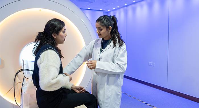
Ambient MRI
● Cutting-Edge Technology: Experience the latest Philips Ambient MRI for immersive and relaxing scanning.
● Expertise: Trust our experienced team of radiologists for accurate diagnosis.
● Comfortable Environment: Ambient lighting and visuals reduce claustrophobia, making your MRI stress-free.
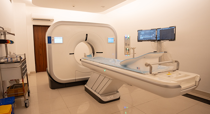
CT Scan
● Cross-Sectional Imaging: Provides detailed slice-by-slice images for accurate diagnosis of internal anatomy and conditions.
● Advanced Diagnostic Applications: Widely used for cancer detection, lung analysis, angiographic studies, and radiotherapy planning.
● Revolutionary Technology: Features innovative advancements like 128 slices/second reconstruction for faster, precise results.
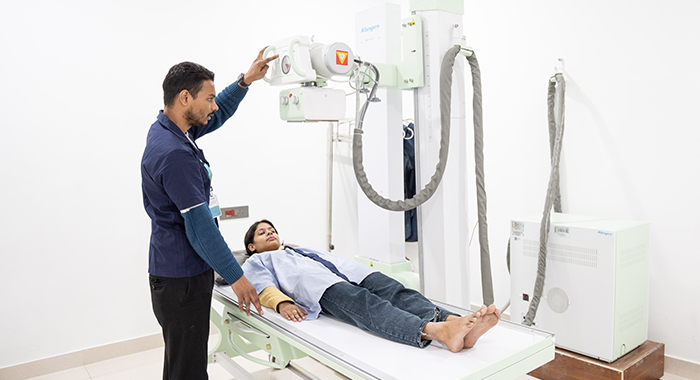
Digital X-RAY
● Advanced Imaging Technology: Captures X-ray images on specialised cassettes, processed digitally for enhanced clarity and precision.
● Efficient and Safe: Offers immediate image preview, eliminates chemical processing, and uses less radiation compared to conventional methods.
● Enhanced Diagnostic Accuracy: Wider dynamic range and special image processing techniques improve image quality, aiding in accurate diagnosis.
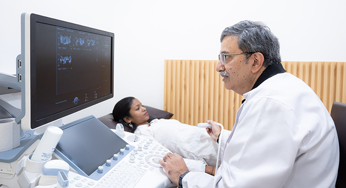
Ultrasound
● Comprehensive Imaging: Utilises Dual-Energy X-Ray Absorptiometry (DEXA) for precise bone mineral density measurement.
● Non-invasive and Painless: Uses high-frequency sound waves to create safe and comfortable imaging without invasive procedures.
● Versatile Diagnostic Tool: Applicable for examining the abdomen, fetus, and subcutaneous tissues to identify potential diseases or lesions.
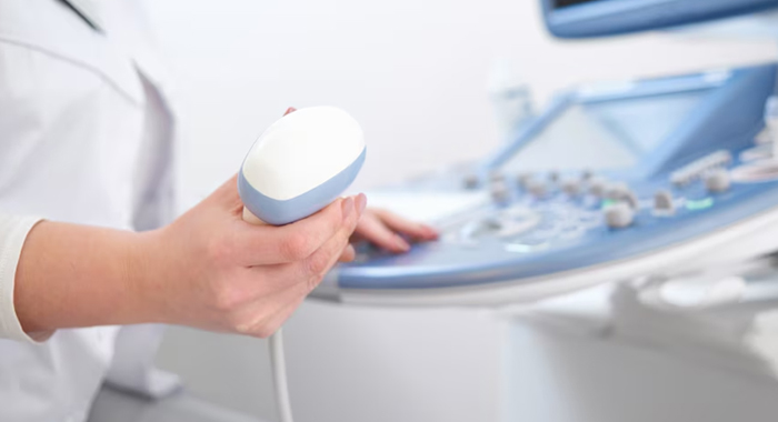
Color Doppler
● Blood Flow Assessment: Evaluates blood flow in major arteries and veins, identifying blockages, clots, and narrowing for conditions like carotid artery issues or deep vein thrombosis.
● Pregnancy Monitoring: Used during pregnancy to check blood flow in the fetus, ensuring the baby's health.
● Targeted Varicose Vein Analysis: Assesses vein valve competency and perforators for precise varicose vein treatment.
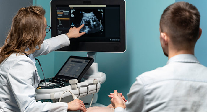
Echocardiography
● Comprehensive Heart Imaging: Echo creates moving pictures of the heart to assess its size, shape, chamber functionality, and valve performance.
● Diagnostic Applications: Detects poor blood flow, previous heart attack injuries, blood clots, fluid build-up, and issues with major blood vessels. It is also useful for diagnosing heart problems in infants and children.
● Additional Insights: Measures heart dimensions, cardiac output, and ejection fraction for a detailed evaluation. Prior appointment is recommended.
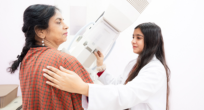
Mammography
● Early Breast Cancer Detection: Mammography uses low-dose X-rays to detect breast cancer early, with screenings recommended every 2 years for women aged 50-74 or after 35 for high-risk individuals.
● Complementary Imaging Techniques: Ultrasound (sonomammography) is used to evaluate masses found on mammograms or for young women with dense glandular breast tissue.
● Comprehensive Screening Approach: Combines mammography with physical breast examinations to provide an effective method for detecting early-stage breast cancer.

CBCT
● Comprehensive 3D Imaging: Captures detailed visuals of bone structure, soft tissues, and nerve pathways, aiding in precise dental implant planning and diagnosis.
● Innovative Technology: Uses a cone-shaped radiation source and advanced detectors for high-resolution, low-radiation imaging, ensuring safety and accuracy.
● Advanced Features: Equipped with movement artefact correction, ultra-low dose imaging, and endodontic mode for capturing fine details in complex dental cases.
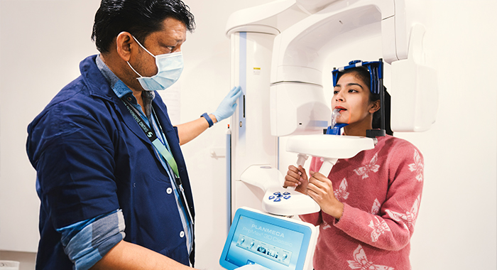
OPG & Cephalogram
● Comprehensive Dental Imaging: OPG (Orthopantomogram) provides a panoramic view of the entire mouth, including teeth, jaw, and surrounding structures, ensuring detailed diagnostics.
● Precise Orthodontic Analysis: Cephalogram offers a lateral view of the skull, enabling accurate assessment of jaw alignment and craniofacial relationships for orthodontic treatments.
● Non-Invasive and Painless: OPG and Cephalogram are safe, painless imaging techniques designed for quick and effective dental and orthodontic care diagnosis.
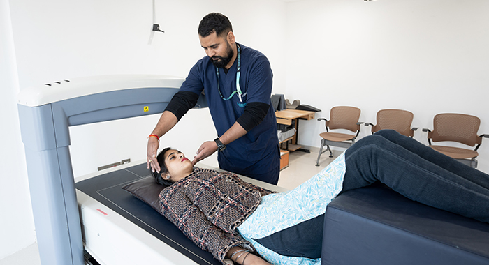
DEXA Scan
Dual-Energy X-Ray Absorptiometry (DEXA) is the gold standard in assessing bone mineral density (BMD), essential for diagnosing osteoporosis, predicting fracture risk, and evaluating body composition accurately.
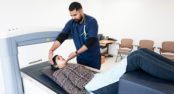
Bone Densitometry
● Advanced Technology: Utilises Dual-Energy X-Ray Absorptiometry (DEXA) for precise bone mineral density measurement.
● Low Radiation Exposure: Employs minimal radiation, less than 1/20th of a chest X-ray, ensuring patient safety.
● Early Osteoporosis Detection: Identifies bone loss at initial stages, facilitating timely intervention and treatment.

Hysterosalpingogram
● Purpose and Benefits: HSG is an X-ray test to examine the uterus and fallopian tubes, commonly used in fertility treatments to identify causes of infertility and guide treatment plans.
● Procedure and Timing: Scheduled 2–5 days after your period ends, before ovulation. Patients may experience manageable cramping similar to menstrual pain, with antispasmodics or pain medication provided if needed.
● Safety and Risks: The procedure is generally safe, with rare risks of pelvic infection or allergic reactions to the dye. Antibiotics are prescribed in the rare event of complications.

Amniocentesis
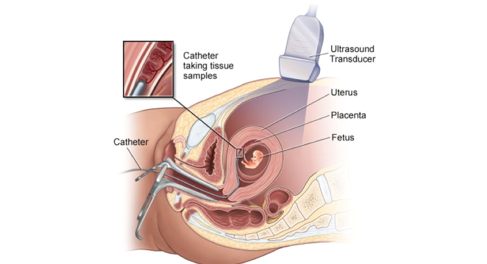
● Diagnostic Procedure: Amniocentesis is a prenatal test that involves extracting a small sample of amniotic fluid to assess the health of the baby and detect genetic or chromosomal conditions.
● Comprehensive Testing: Helps diagnose conditions such as Down syndrome, neural tube defects, and other genetic abnormalities, offering valuable insights into the baby’s development.
● Safe and Guided: Performed with ultrasound guidance to ensure accuracy and minimise risks, making it a reliable choice for prenatal diagnostics.

