MRI for Cancer Detection: How Indian Oncologists Use It in Treatment Planning

“Hmm, cancer is a big word, isn’t it?” That’s what one of our patients murmured during her MRI scan at Sikund Diagnostic Centre last week. “Aah, it’s scary, but I’m glad there’s something like MRI to help catch it early.”
Yes, exactly. Cancer is a major concern not only globally but also in India, where early detection and precise staging can mean the difference in treatment outcomes. And that’s where MRI comes in, not only as a diagnostic tool, but also as a planning tool for oncologists.
At Sikund Diagnostic Centre, we view MRI as more than just a machine. It’s a non-invasive, highly complex, and really patient-friendly instrument that helps decipher the mysteries of the body without using a scalpel or causing pain.
Let us walk you through how oncologists across India are relying on MRI, particularly for cancers such as prostate, breast, brain, and liver, and how Sikund ensures that your MRI experience is as pleasant and comfortable as possible.
Why MRI? And Why Now?
Okay, first things first: MRI, or Magnetic Resonance Imaging, creates detailed images of your inside structures by using powerful magnetic fields and radio waves. No radioactivity.
Non-invasive. Safe. Particularly effective in soft tissue imaging, where malignancies frequently go undetected.
Now, let’s go into specifics.

Prostate Cancer
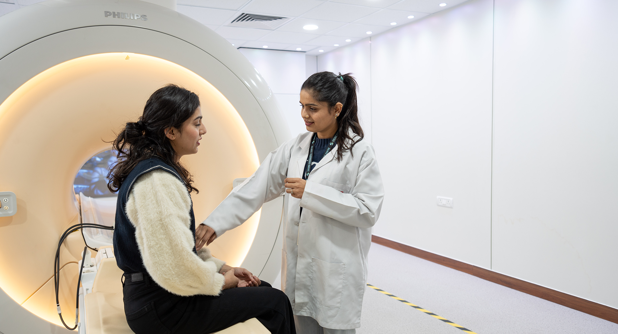
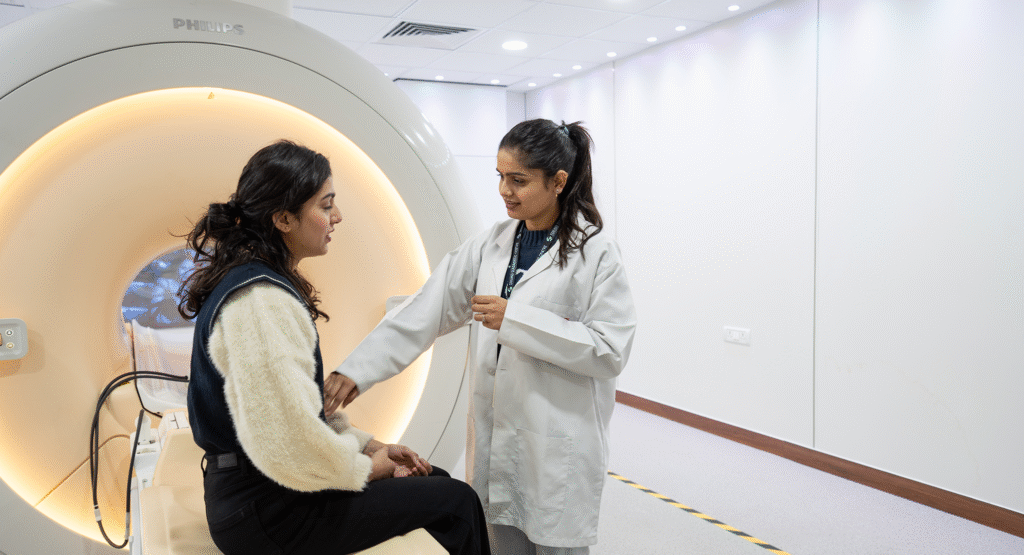
"This test is for prostate cancer, correct?" But it doesn't hurt?" one of our male patients recently inquired. Right, the MRI does not hurt at all. Multi-parametric MRI (mpMRI) has revolutionised prostate cancer detection. This test assesses the prostate from various perspectives using T2-weighted images, diffusion-weighted imaging (DWI), and dynamic contrast enhancement (DCE), resulting in a clear, layered picture. Oncologists use this to detect clinically significant tumours, avoid unnecessary biopsies, and plan focused biopsies or treatments (e.g., focal therapy). At Sikund, our Philips dStream MRI system increases the Signal-to-Noise Ratio (SNR) by up to 40%, resulting in better prostate pictures and more accurate cancer diagnosis and grading.

Breast Cancer
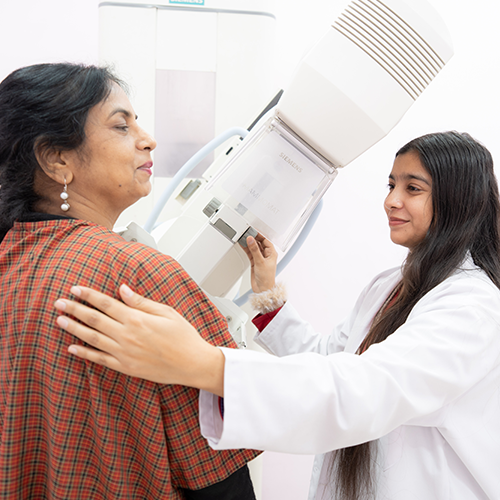
Ah, breast cancer. This is the second most frequent cancer among Indian women. We frequently visit patients who have already had a mammogram or ultrasound but are still seeking answers. This is where contrast-enhanced breast MRI excels. MRI can reveal abnormalities that are too tiny or obscured by mammography, particularly in women with thick breast tissue. Surgeons and oncologists employ MRI to assess tumour size and distribution, detect multicentric or contralateral illness, and monitor chemotherapy responses. "Hmm, this scan feels more peaceful than I expected," a young woman remarked to us as she slept peacefully beneath our ambient lights. We smiled and said, "That's intentional!" Our built-in television, adjustable lights, and personalised sound system are meant to relieve stress and claustrophobia, which we take very seriously, especially for sensitive scans like breast MRIs.
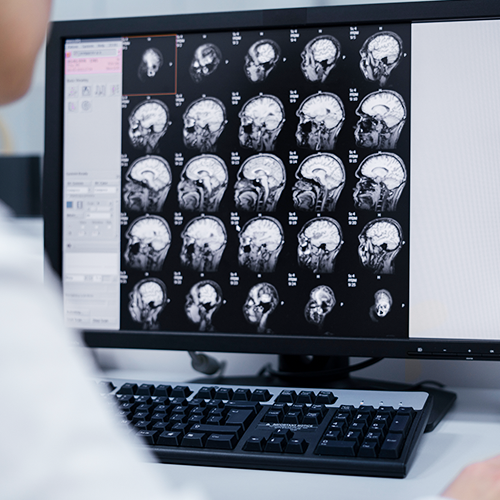
Brain Tumors
Most MRIs do not require fasting. However, if you are having a contrast-enhanced MRI (usually for the abdomen or pelvis), we will ask you to fast for around 4-6 hours. Do not fear, we will confirm this with you when you arrange your appointment.
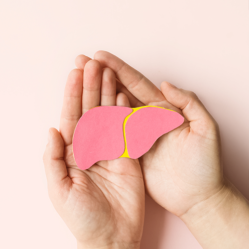
Liver Cancer
Hepatologists and GI oncologists rely on liver MRI. MRI uses fat quantification and contrast-enhanced sequences to evaluate primary liver cancers (such as HCC) and metastases. What sets MRI apart is its ability to distinguish benign tumours (such as hemangiomas or cysts) from cancers, typically without the need for a sample. Oh, and are there any metal implants in your body? Our technique includes MAR (Metal Artefact Reduction), which reduces picture distortions that used to complicate liver MRIs in such circumstances.
How Indian Oncologists Use This Information
“So, once the MRI is done, what happens next?” Great question! Oncologists usually use MRI data to arrange treatment as follows:
- Stages of Cancer
Determining the extent of cancer spread is crucial for deciding whether to undergo surgery, chemotherapy, or radiation. - Pre-surgical Planning
Knowing the exact location and margins of the tumour allows surgeons to avoid injuring neighbouring tissues. - Therapy Monitoring
MRI measures tumour shrinking following chemotherapy or radiation. Oncologists seek functional rather than structural alterations. - Post-treatment surveillance
Hmm, how will we know if cancer has returned after treatment? You guessed it: MRI helps detect recurrences early and safely.


Why Sikund Diagnostic Centre?
Okay, let’s talk about you, the patient who walks into Sikund for an MRI. Whether you’re seven or seventy-seven, we treat patients of all ages, from pediatric to geriatric, with compassion and technological expertise.
Here’s what separates us:
- Ambient MRI Experience: Relax with tailored music or watch your favorite show while lying within the scanner.
- Philips dStream MRI offers industry-leading technology for faster scans with improved resolution.
- Improved SNR by 40%, resulting in more detailed images and fewer repeat scans.
- Features for reducing claustrophobia include adjustable lights, a built-in television, and two-way communication.
- Expert radiologists: Our in-house staff includes oncology-trained radiologists and consultants. Yes, these are the ones your oncologist will contact after reviewing our report.
Final Thoughts
We understand that receiving a cancer diagnosis, or even waiting to rule it out, is stressful. But MRI can provide a sign of comfort by revealing clear, correct information.
“Umm, I didn’t expect it to be this smooth,” commented an elderly guy last week following his liver MRI.
We smiled and said, “That’s what we strive for.”
So, whether it’s staging a tumour, directing a biopsy, or verifying remission, MRI is quietly altering the path of cancer treatment in India.
And we at Sikund Diagnostic Centre are honoured to be a part of that journey, both with and for you.